- Comprehensive cardiac model
- High purity and functionality
- Supports co-culture applications to study cell interactions within the cardiac microenvironment
- Enables advanced toxicity and drug screening studies
.png)
Cardiotoxicity Testing and Drug Screening
Ncyte® Heart in a Box™ offers a comprehensive platform for assessing the cardiotoxic effects of potential drug candidates. By incorporating cardiomyocytes, endothelial cells, and cardiac fibroblasts, this system allows researchers to evaluate drug effects on cardiac function, viability, and toxicity across various heart cell types. High-throughput screening and large-scale compound testing can predict cardiotoxicity risks before clinical trials, reducing the reliance on animal models.
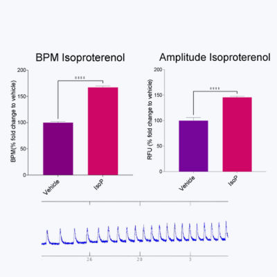 A. Response of 3D Cardiac Microtissues to tool compounds
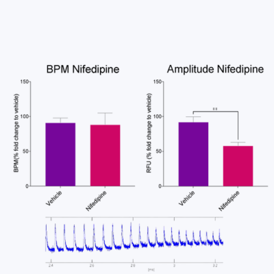 B: Response of 3D Cardiac Microtissues to tool compounds
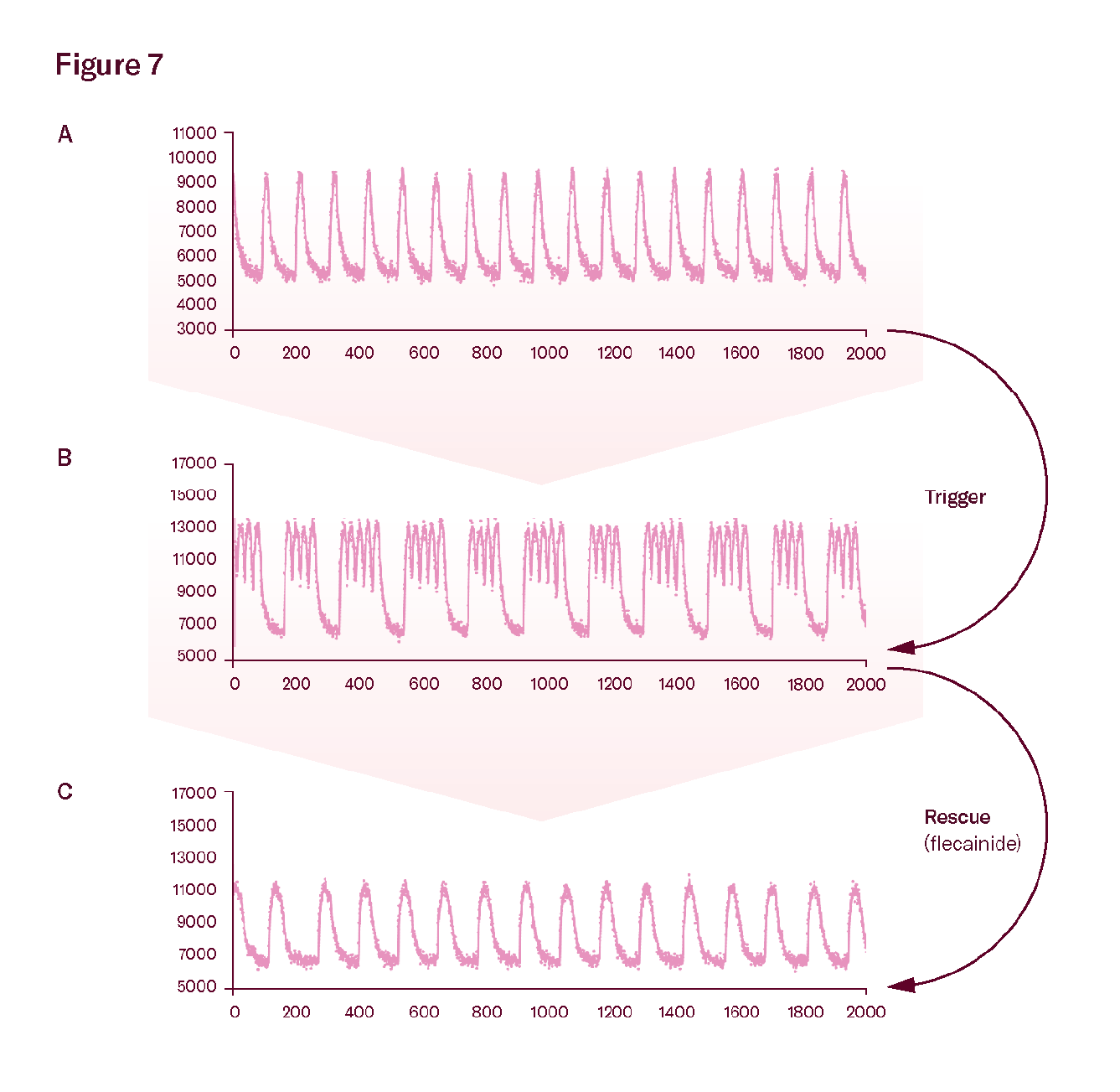 Disease modeling with Ncardia’s cardiac microtissues: CPVT.
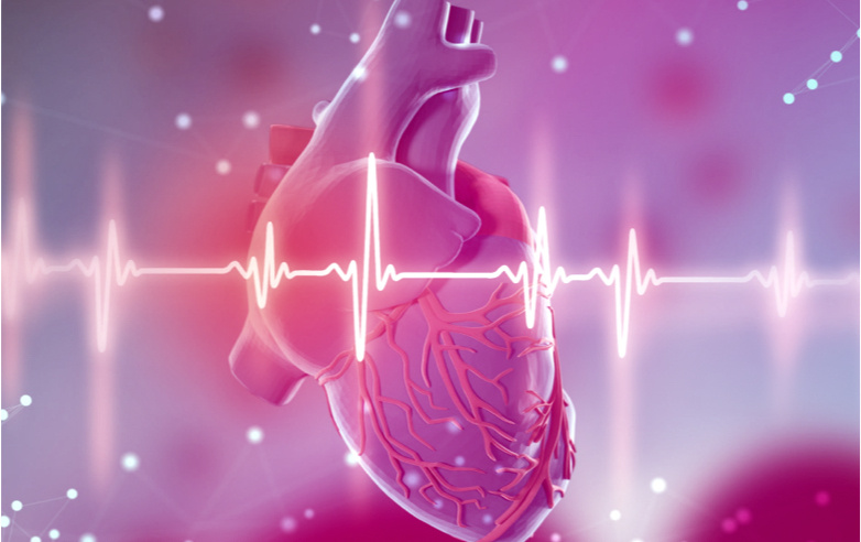
Heart Disease Modeling
This system is ideal for modelling various
cardiovascular diseases, including ischemic
heart disease, heart failure, hypertrophy, and
fibrosis. Researchers can explore disease
mechanisms in a human-relevant 3D envi-
ronment, gaining a deeper understanding of
cellular interactions and disease progression.
Ncyte® Heart in a Box™ is particularly useful
for studying how cardiomyocytes, endothelial
cells, and fibroblasts interact during heart
disease development.
.png) Response to Drugs in the Heart in a Box Model
Response to Drugs in the Heart in a Box Model
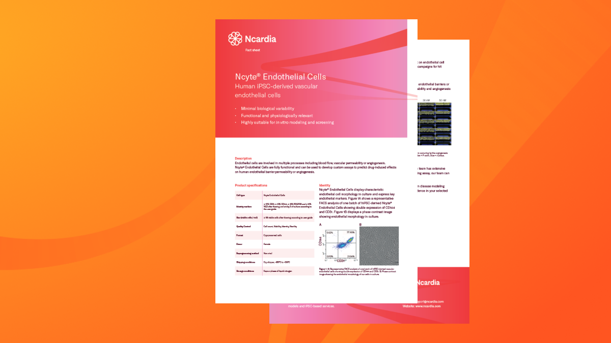
Angiogenesis and Vascular Remodeling
Ncyte® Endothelial Cells form functional
vascular networks, providing an excellent
model for studying angiogenesis and vascular
remodelling, especially in ischemic heart
diseases. This application is valuable for
testing therapies aimed at promoting blood
vessel growth and improving circulation,
such as in post-myocardial infarction or
heart failure.
.png) Ac-LDL uptake assay showing Ncyte® Endothelial Cells’ ability to internalize acetylated low-density lipoprotein
Ac-LDL uptake assay showing Ncyte® Endothelial Cells’ ability to internalize acetylated low-density lipoprotein
Fibrosis Research and ECM Remodeling
Cardiac fibrosis, a hallmark of many heart
diseases, is driven by the activation of car-
diac fibroblasts. Ncyte® Cardiac Fibroblasts
offer a unique opportunity to study ECM re-
modelling, collagen deposition, and fibroblast
activation in response to injury. This system
is critical for developing antifibrotic therapies
to reduce scar tissue formation and restore
heart function.
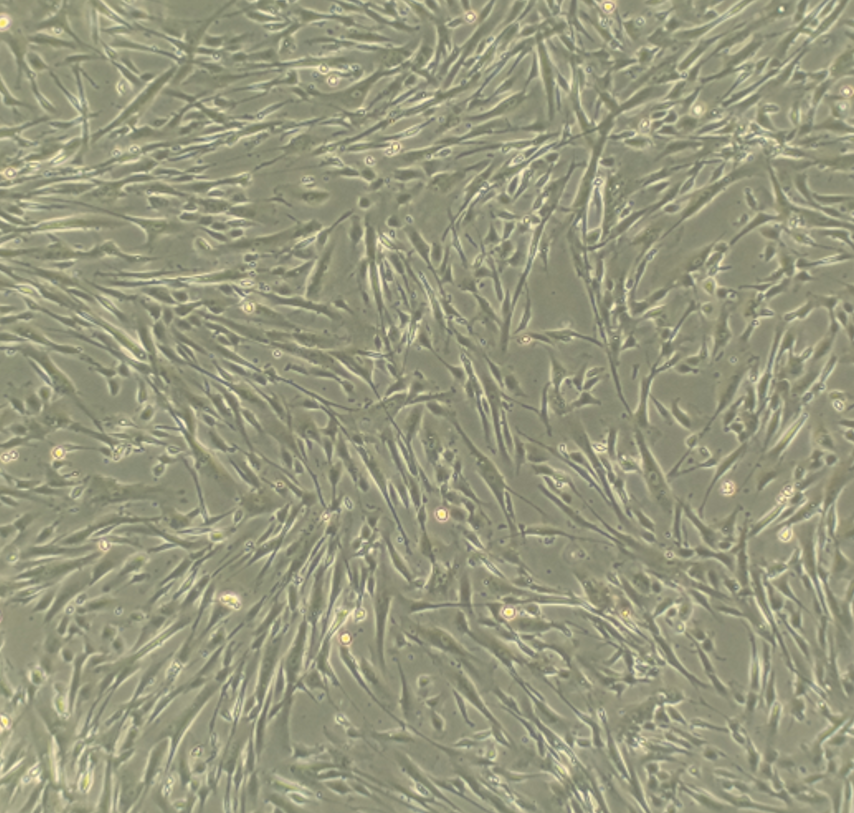 Brightfield image of Ncyte® cardiac fibroblasts showing their elongated and spindle-shaped morphology
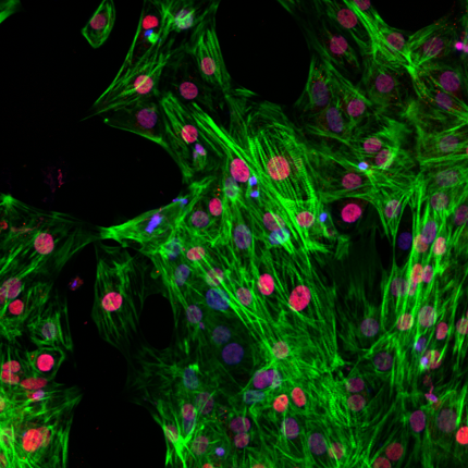
.png) Immunofluorescence staining for vimentin (green) and connexin 43 (CX43, red). Vimentin is a cytoskeletal protein present in fibroblasts, while CX43 is a gap junction protein involved in cell-to-cell communication. DAPI (blue) stains the cell nuclei. Scale bar: 50 μm
Immunofluorescence staining for vimentin (green) and connexin 43 (CX43, red). Vimentin is a cytoskeletal protein present in fibroblasts, while CX43 is a gap junction protein involved in cell-to-cell communication. DAPI (blue) stains the cell nuclei. Scale bar: 50 μm
.png) Immunofluorescence staining for collagen type I (COL1A1, red) and CX43 (green). COL1A1 is a major component of the extracellular matrix. DAPI (blue) stains the cell nuclei.
Immunofluorescence staining for collagen type I (COL1A1, red) and CX43 (green). COL1A1 is a major component of the extracellular matrix. DAPI (blue) stains the cell nuclei.
Human iPSC-derived atrial cardiomyocytes
Neural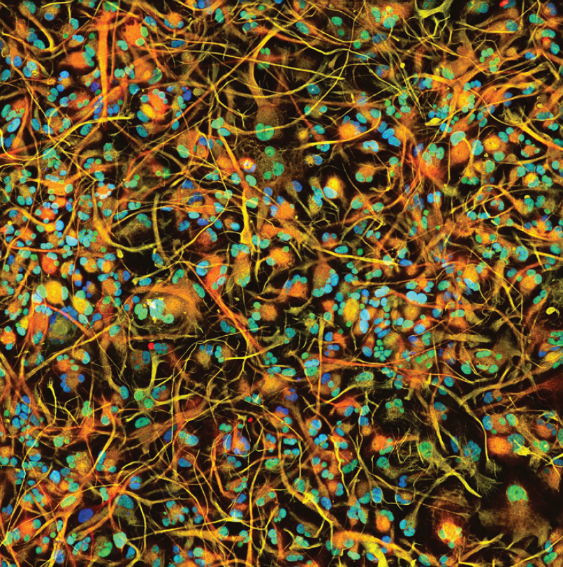
Human iPSC-derived astrocytes
Vascular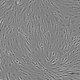
Human iPSC-derived vascular endothelial cells
CardiacHuman iPSC-derived 3D cardiac microtissue model
Neural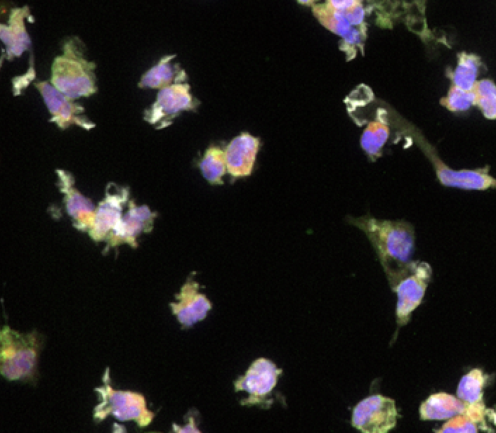
Human iPSC-derived microglia
Cardiac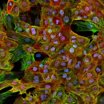
Non-Human Primate Cynomolgus iPSC-Derived Ventricular-Like Cardiomyocytes
Vascular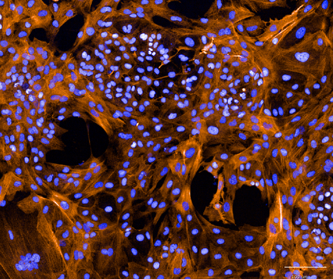
Human iPSC-derived vascular smooth muscle cells
Cardiac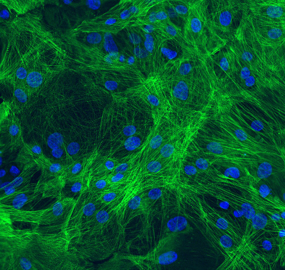
Human iPSC-derived ventricular cardiomyocytes