Order Now
Benefits
- Rapidly evaluate therapeutic compounds
- Effectively mediate inflammatory responses
- Valuable for investigating cytokine release in neuroinflammatory conditions
neural
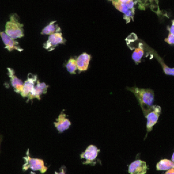
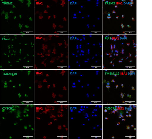
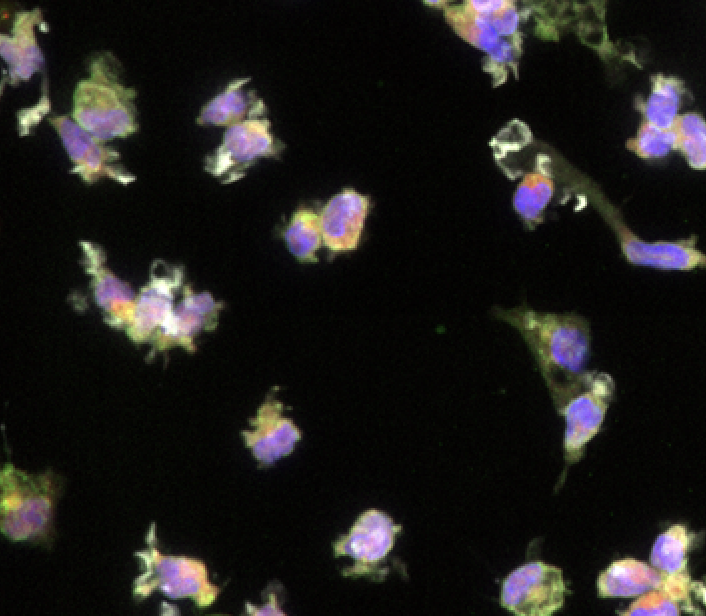
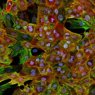
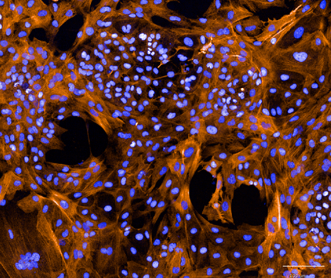
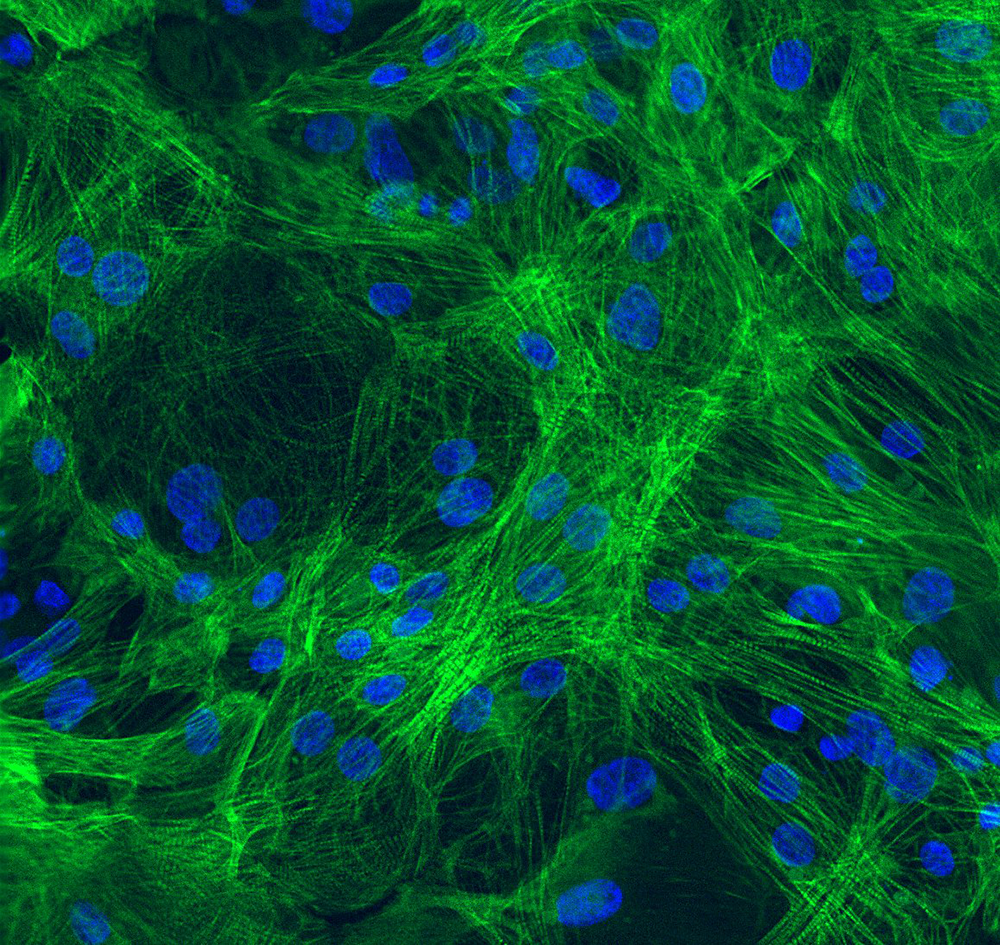
- Immunofluorescence staining demonstrates robust expression of microglial markers including TREM2, IBA1, PU.1, TMEM119, and CX3CR1 in Ncyte® Microglia.
- Confocal microscopy reveals co-localization of microglial markers, indicating the presence of a mature microglial phenotype.
- Ncyte® Microglia exhibit characteristic ramified morphology, further supporting their microglial identity.
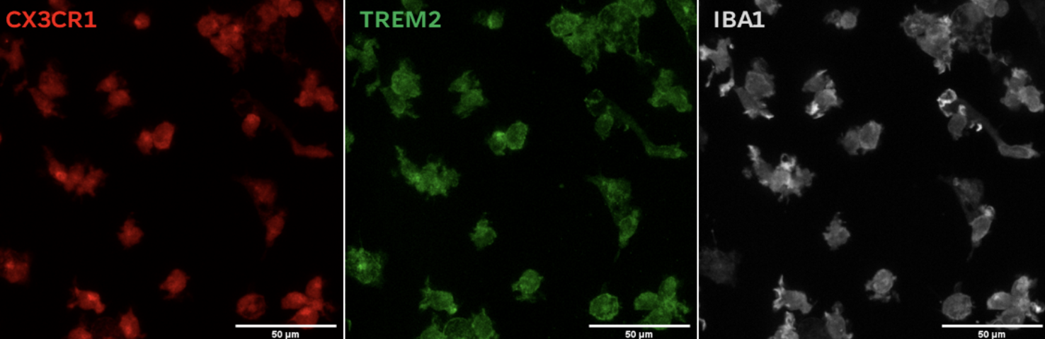 Triple positive population for CX3CR1, TREM2, and IBA1 indicates mature microglial phenotype.
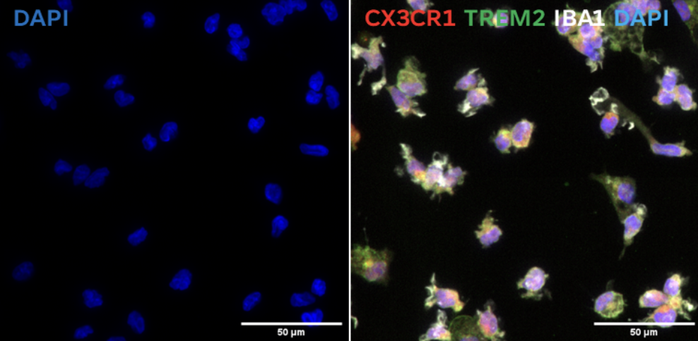 Expression of CX3CR1 and TREM2 suggests suitability for co-culture systems with neurons to model Alzheimer's Disease and other neurodegenerative diseases.
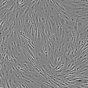
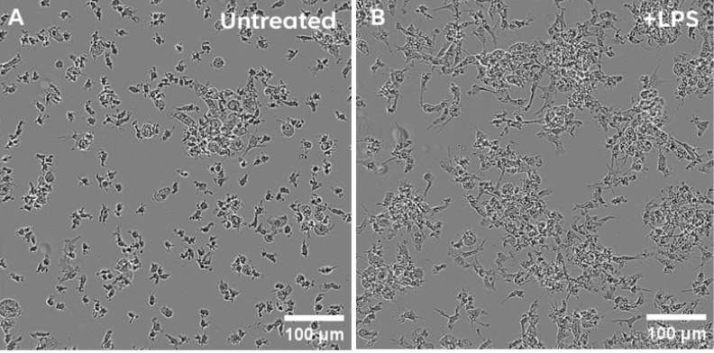 Brightfield images of Ncyte® Microglia on day 7 post-thaw. A) Untreated microglia and B) Microglia treated with LPS (100 ng/ml) for 18hrs.
- Morphological evidence of activation is demonstrated by changes in Ncyte® Microglia morphology following LPS stimulation.
- Flow cytometry and ICC confirm robust expression of key microglial markers (CX3CR1, TREM2, IBA1, CD45, CD11b) in both resting and activated states.
- Ncyte® Microglia show significant cytokine release upon LPS stimulation, highlighting their functional role in mediating inflammatory responses.
- Ncyte® Microglia offer an ideal in vitro model for studying neuroinflammation and the mechanisms of neurodegenerative diseases, including
Alzheimer’s and Parkinson’s.
- Ncyte® Microglia exhibit a strong pro-inflammatory reaction upon LPS stimulation.
- LPS treatment significantly increases the production of IL-6 and TNF-α, key inflammatory mediators. These results underscore the ability of Ncyte® Microglia to effectively mediate inflammatory responses.
- Ncyte® Microglia provide a valuable in vitro model for investigating cytokine release in neuroinflammatory conditions.

Cardiac
 Ncyte® aCardiomyocytes
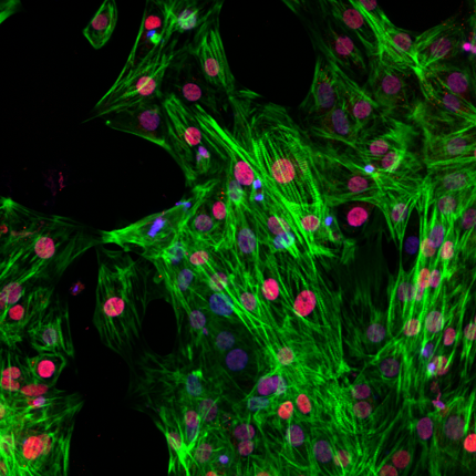
 Ncyte® Astrocytes
 Ncyte® Endothelial Cells
.png) Ncyte® Heart in a Box™
Ncyte® Heart in a Box™
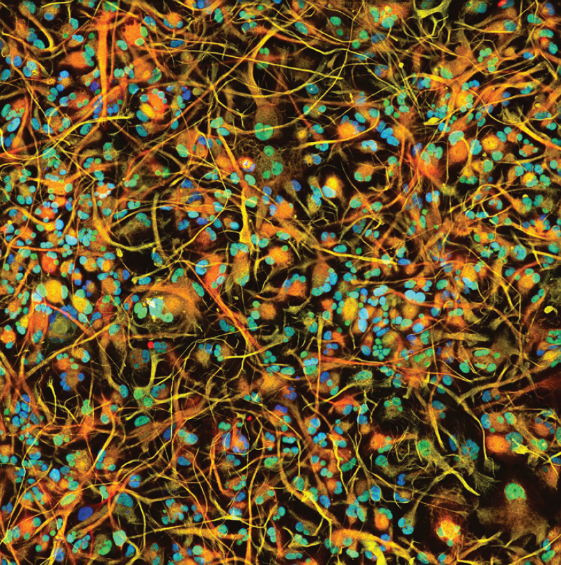
 Ncyte® Microglia
 Ncyte® NHP-C vCardiomyocytes
 Ncyte® Smooth Muscle Cells
 Ncyte® vCardiomyocytes
Human iPSC-derived atrial cardiomyocytes
NeuralHuman iPSC-derived astrocytes
VascularHuman iPSC-derived vascular endothelial cells
CardiacHuman iPSC-derived 3D cardiac microtissue model
NeuralHuman iPSC-derived microglia
CardiacNon-Human Primate Cynomolgus iPSC-Derived Ventricular-Like Cardiomyocytes
VascularHuman iPSC-derived vascular smooth muscle cells
CardiacHuman iPSC-derived ventricular cardiomyocytes