- High purity ventricular-like cardiomyocytes
- Assay-ready format: no pre-culture or dissociation required
- Visible beating clusters within 48 hours of seeding
- Fully functional and electrophysiologically active
- Ideal for electrophysiology and cardiotoxicity testing
Ncyte® Plate-Ready vCardiomyocytes are characterized by flow cytometry to ensure a purity of ≥ 70% cardiac Troponin T (cTnT) positive cells right after thawing (Figure 1). They typically also express Myosin Light Chain 2v (MLC2v), indicating a ventricular-like phenotype.
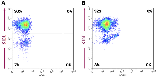 Figure 1. A) Flow cytometry analysis of a representative batch showing cTnT positive cells (91.5%) and cTnT/MLC2v double positive cells (89.27%).
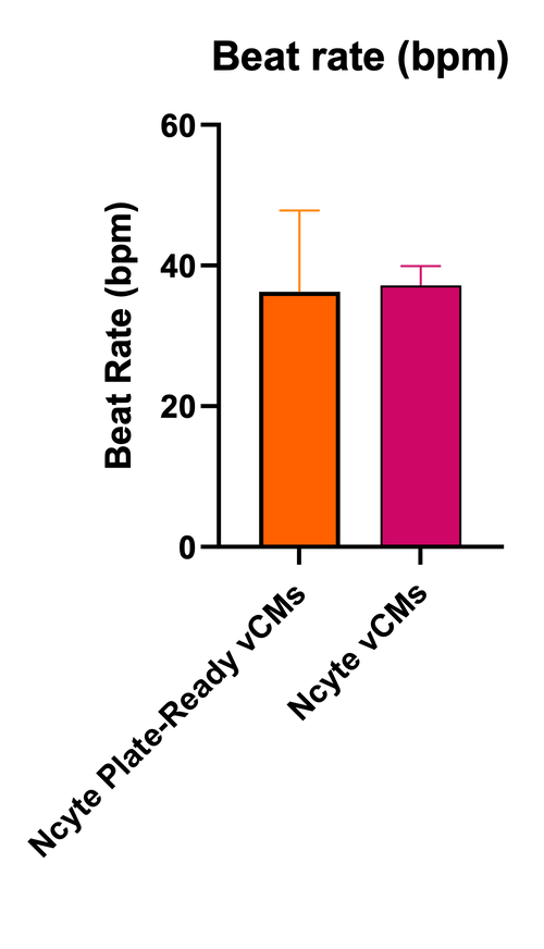
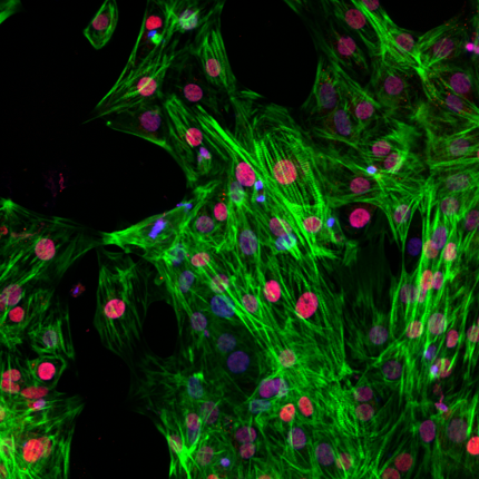
Ncyte® Plate-Ready vCardiomyocytes are a physiologically relevant model for the phenotypic study of cardiac diseases. They feature well aligned myofibrils and intact structural sarcomere organization (Figure 2), enabling robust pacing and electrophysiological readouts. In addition, Ncyte® Plate-Ready vCardiomyocytes present a robust electrode coverage (Figure 3A) and a relatively slow and uniform beating rate, similar to the Ncyte® vCardiomyocytes (Figure 3B).
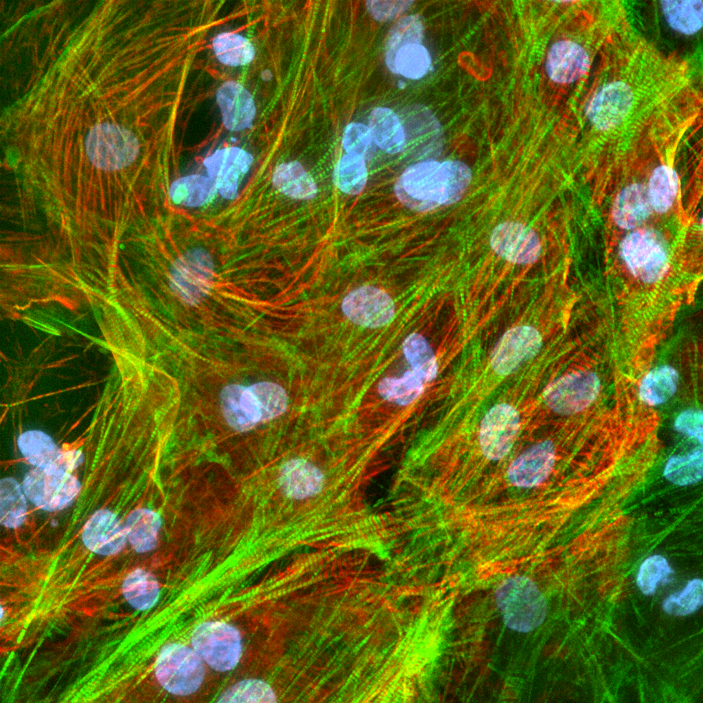
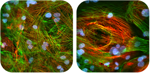 Figure 2. Immunofluorescence staining of Ncyte® Plate-ready vCardiomyocytes showing Cardiac Troponin T (Green), MLC2V (Red) and DAPI (Blue). 60x magnification.
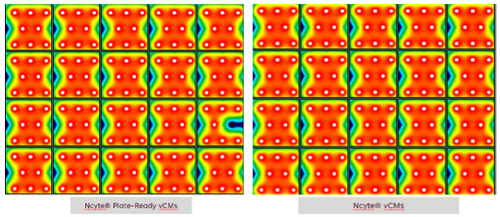 Figure 3. A) Microelectrode array baseline beat rate of a 96-well plate with Ncyte® Plate-Ready vCardiomyocytes and Ncyte® vCardiomyocytes.
These cells are highly responsive to pharmacological modulation such as adrenergic stimulation with isoproterenol and to ion channel blockers such as nifedipine (L-type calcium channel blocker) and dofetilide (hERG channel blocker; Figure 4A–C). Thus, they can be utilized in functional assays including imaging, impedance, capacitance, and contractility, metabolic and cell viability assays, as well as for biomarker identification.
Leverage Ncardia’s custom iPSC-based services—from disease modeling to in vitro pharmacology and cardiotoxicity screening — to de-risk candidate selection and advance your drug discovery pipeline. Combine Ncyte® Plate-Ready vCardiomyocytes with our validated cardiotoxicity assays for a comprehensive evaluation of cardiac safety.
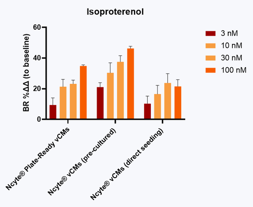 Figure 4. A) Acute exposure to isoproterenol increased the beat rate of Ncyte® Plate-Ready vCardiomyocytes, as seen alongside pre-cultured Ncyte® Cardiomyocytes and directly seeded Ncyte® vCardiomyocytes. Data presented as mean ±SD (n = 3) of the percentage change to baseline recordings for each well.
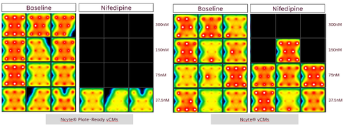 B) A 30-minute exposure to nifedipine, an L-type calcium channel blocker, causes beating arrest in Ncyte® Plate-Ready vCardiomyocytes.
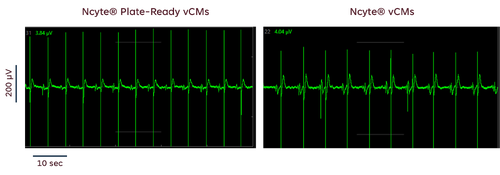 C) Representative MEA traces after 30-minute exposure to dofetilide and an hERG (Ikr) channel blocker, at concentrations ≥ 10 nM, showing notable arrhythmias in Ncyte® Plate-Ready vCardiomyocytes.
Ncyte® Plate-Ready vCardiomyocytes are a versatile and scalable tool for:
- Cardiotoxicity screening
- Electrophysiological assays (e.g., MEA)
- Drug development (from hit identification to lead optimization)
- Mechanistic and disease modeling studies
- Regenerative medicine research
These cells can replace standard formats in workflows where time, consistency and high-throughput readiness are critical. By skipping the 3-day pre-culture and dissociation step, researchers gain immediate access to a robust and reproducible model of human ventricular cardiomyocytes.
The Plate-Ready format allows direct seeding after thawing — ideal for rapid, high-throughput assays with minimal hands-on time. The Pre-Culture format, which includes a 3-day culture and dissociation step, offers greater flexibility for custom setups or long-term studies. Both formats deliver consistent quality and functional performance, allowing you to choose based on speed or experimental complexity.
Human iPSC-derived atrial cardiomyocytes
Neural
Human iPSC-derived astrocytes
Vascular
Human iPSC-derived vascular endothelial cells
Cardiac.png)
Human iPSC-derived 3D cardiac microtissue model
Neural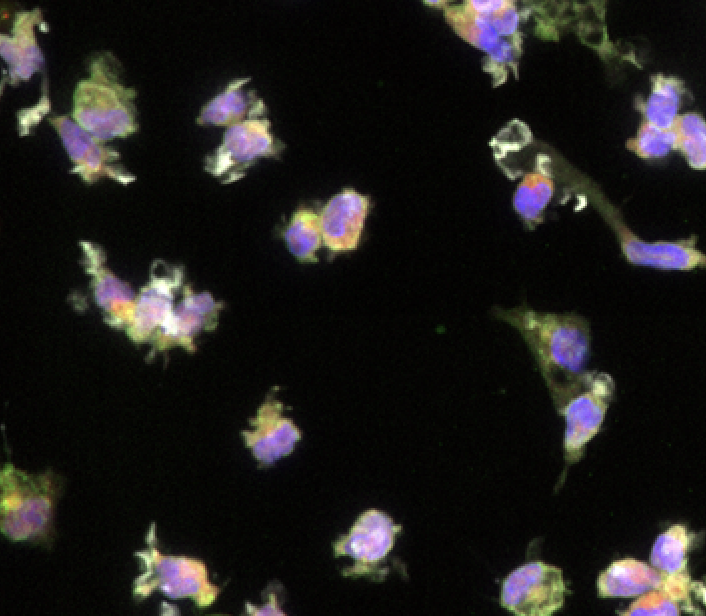
Human iPSC-derived microglia
Cardiac
Non-Human Primate Cynomolgus iPSC-Derived Ventricular-Like Cardiomyocytes
Vascular
Human iPSC-derived vascular smooth muscle cells
Cardiac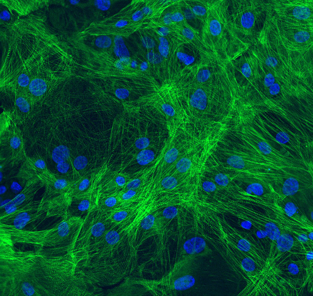
Human iPSC-derived ventricular cardiomyocytes