Filter
Cardiac
 Ncyte® aCardiomyocytes
 Ncyte® Astrocytes
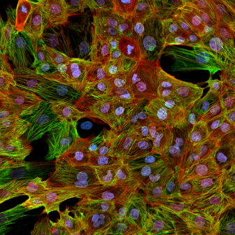
 Ncyte® Endothelial Cells
.png) Ncyte® Heart in a Box™
Ncyte® Heart in a Box™
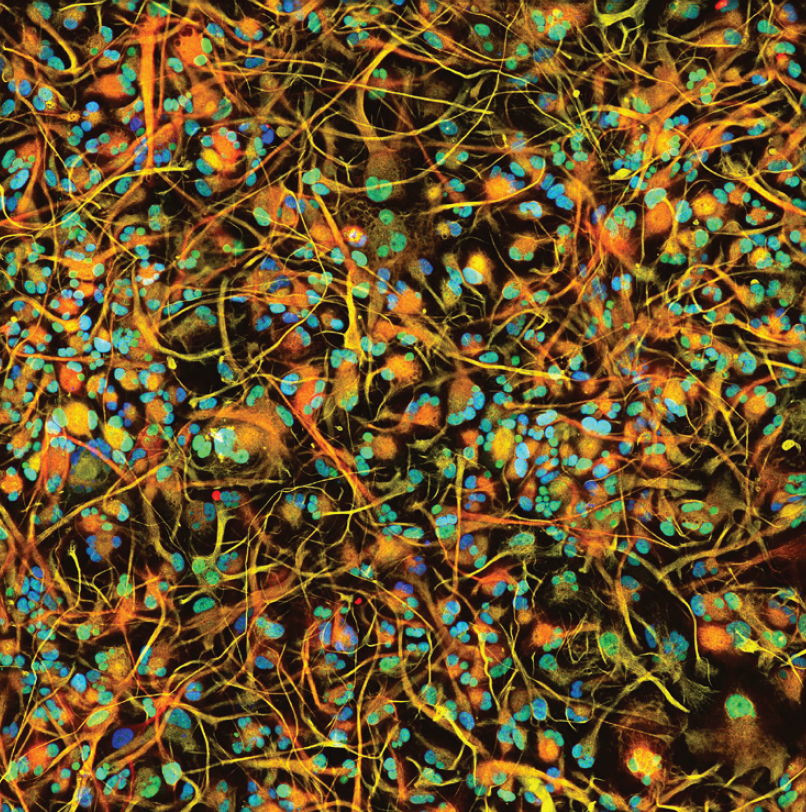
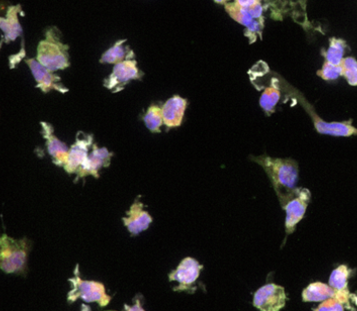 Ncyte® Microglia
 Ncyte® NHP-C vCardiomyocytes
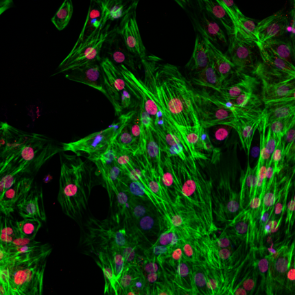
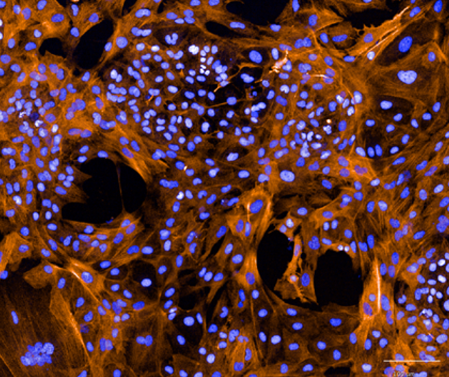 Ncyte® Smooth Muscle Cells
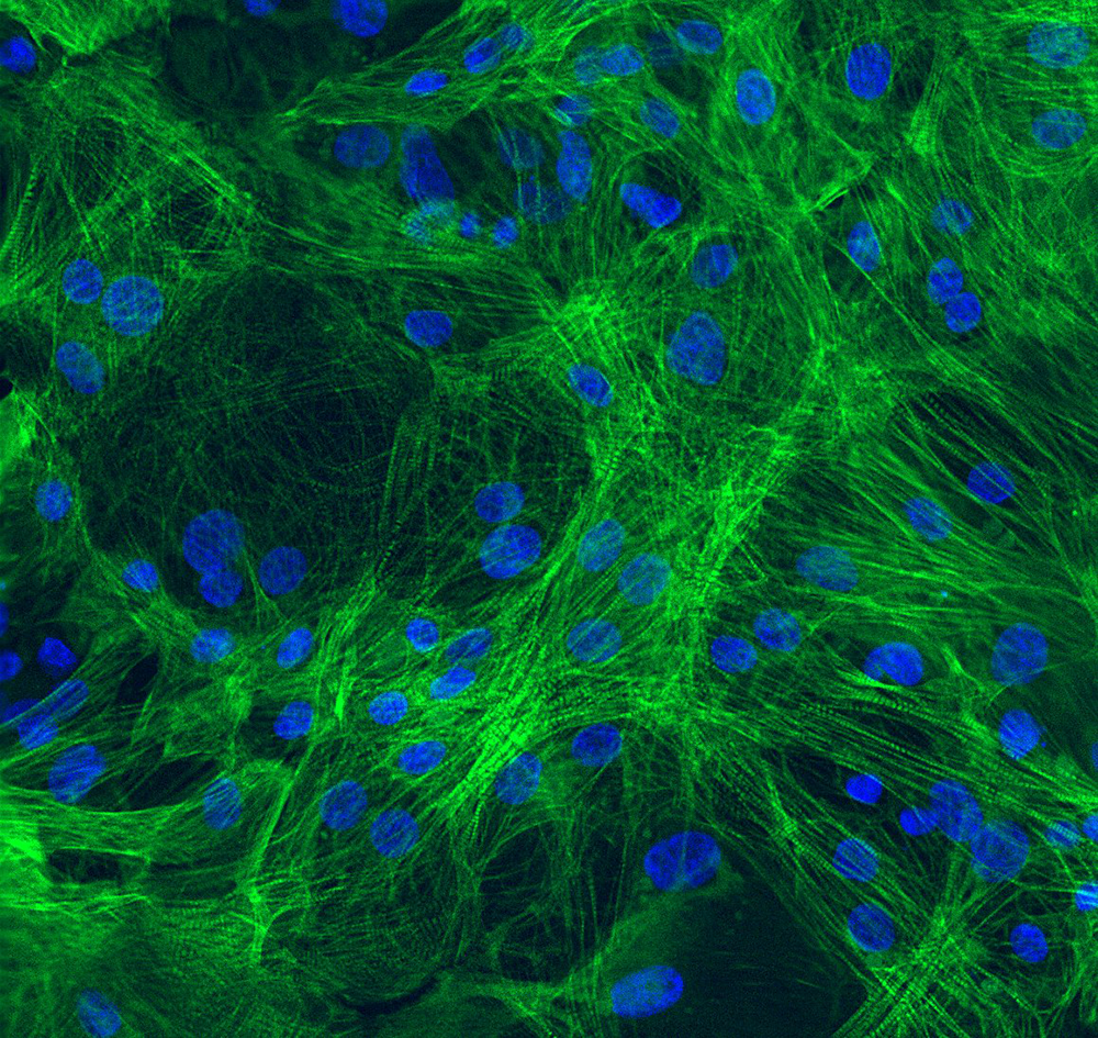 Ncyte® vCardiomyocytes
Human iPSC-derived atrial cardiomyocytes
NeuralHuman iPSC-derived astrocytes
VascularHuman iPSC-derived vascular endothelial cells
CardiacHuman iPSC-derived 3D cardiac microtissue model
NeuralHuman iPSC-derived microglia
CardiacNon-Human Primate Cynomolgus iPSC-Derived Ventricular-Like Cardiomyocytes
VascularHuman iPSC-derived vascular smooth muscle cells
CardiacHuman iPSC-derived ventricular cardiomyocytes

Incorporate relevant cell models
in your research
We’re ready to help make your next step the very best it can be. So let’s start with a conversation – about your vision, goals and expectations for your projects.
Mariana Argenziano, PhD
Associate Director, Manufacturing Technology