- High purity ventricular-like cardiomyocytes
- Physiologically relevant
- Highly suitable for electrophysiological research
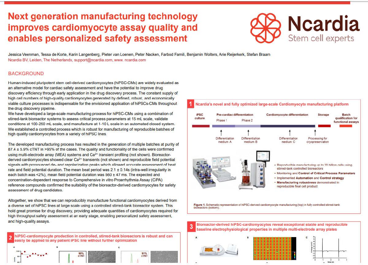
Ncyte® vCardiomyocytes are characterized by flow cytometry to ensure a purity of ≥ 90% cardiac TroponinT (cTnT) positive cells after 3 days in culture following Ncardia’s user guide (Figure 2A). They typically express Myosin Light Chain 2v (MLC2v), indicating a ventricular-like phenotype (Figure 2B). Based on RNA-seq data, Ncyte® vCardiomyocytes express common sarcomere markers, such as TNNT2, while hiPSC and cardiac progenitor markers are silenced. Expression levels of additional genes are available upon request.
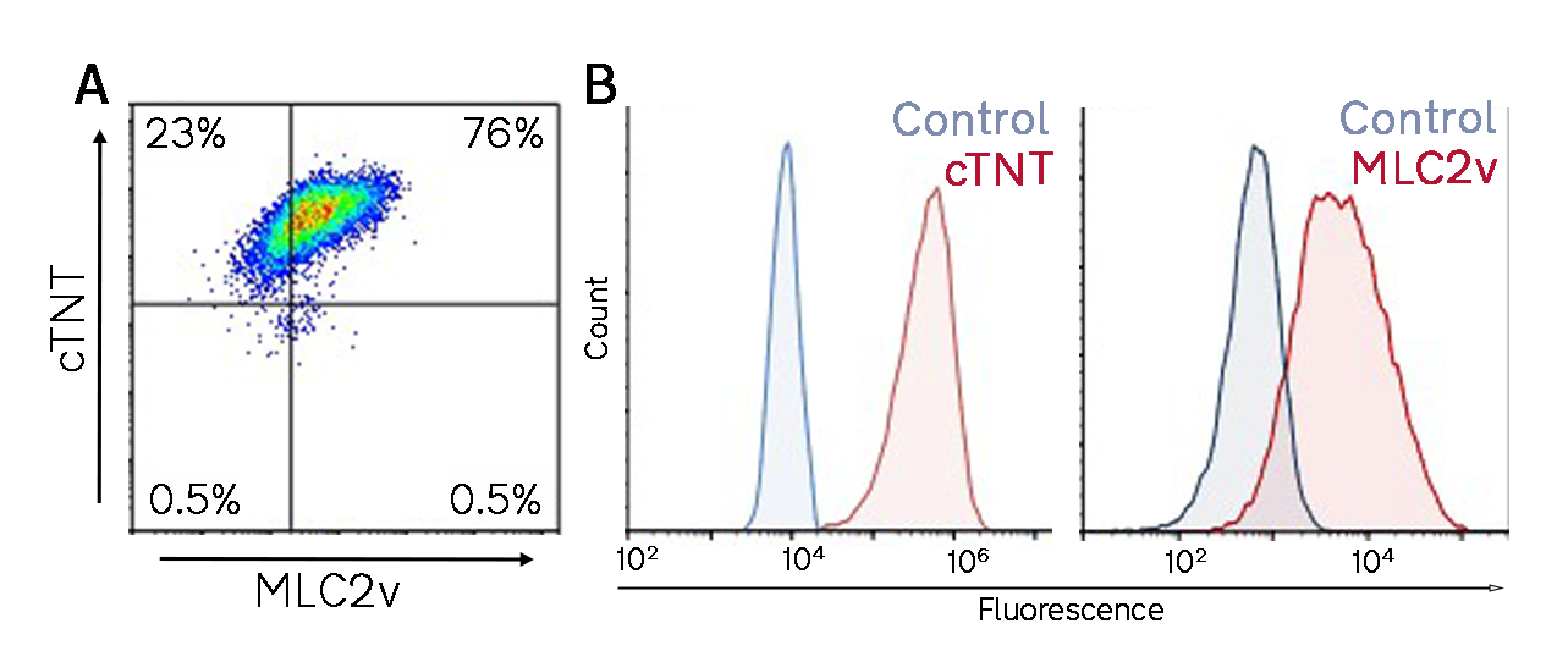 A) Flow cytometry analysis of one representative batch showing cTnT positive cells (99%) and cTnT/MLC2v double positive cells (76%). B) Histograms of cTNT and MLC2v fluorescence signal and their antibody controls.
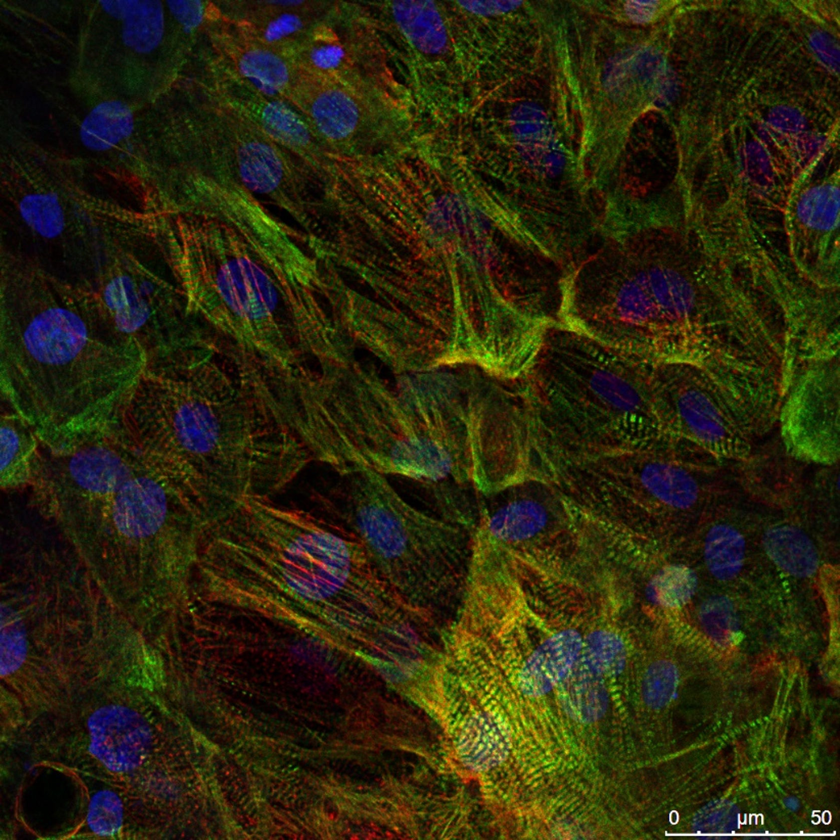
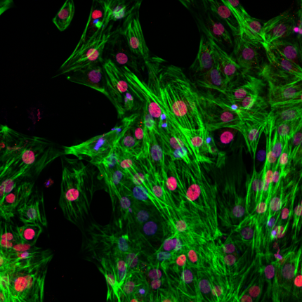
Ncyte® vCardiomyocytes exhibit well aligned myofibrils and intact structural sarcomere organization (See figure A). They typically express Myosin Light Chain 2v (MLC2v), indicating a ventricularlike phenotype. Based on RNA-seq data, Ncyte® vCardiomyocytes express common sarcomere markers, such as TNNT2, while hiPSC and cardiac progenitor markers are silenced. Expression levels of additional genes are available upon request.
Their action potential shows fast upstroke velocity and a well-defined plateau phase, resembling human ventricular cardiomyocytes (See figure B,C).
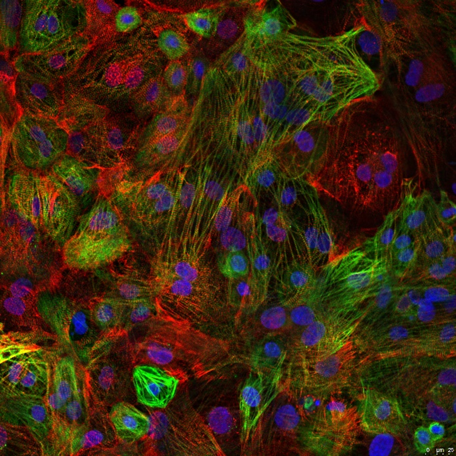 Immunofluorescence staining of Ncyte® vCardiomyocytes: Alpha-actinin (Red), myosin heavy-chain 6 (Green), and DAPI (Blue).
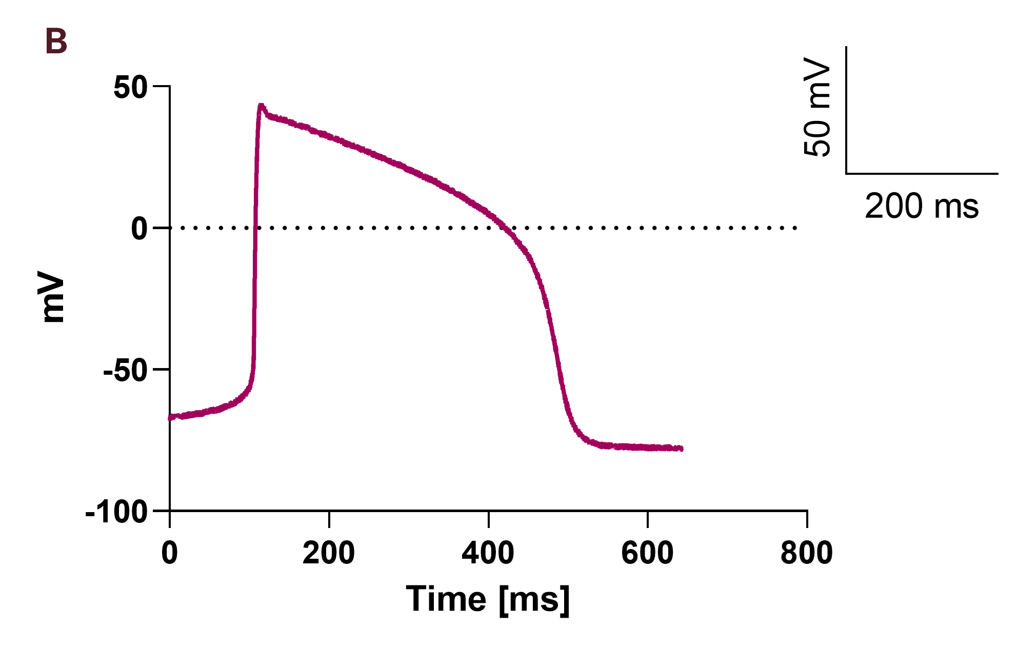 Representative patch-clamp action potential for a batch of Ncyte® vCardiomyocytes in current clamp mode.
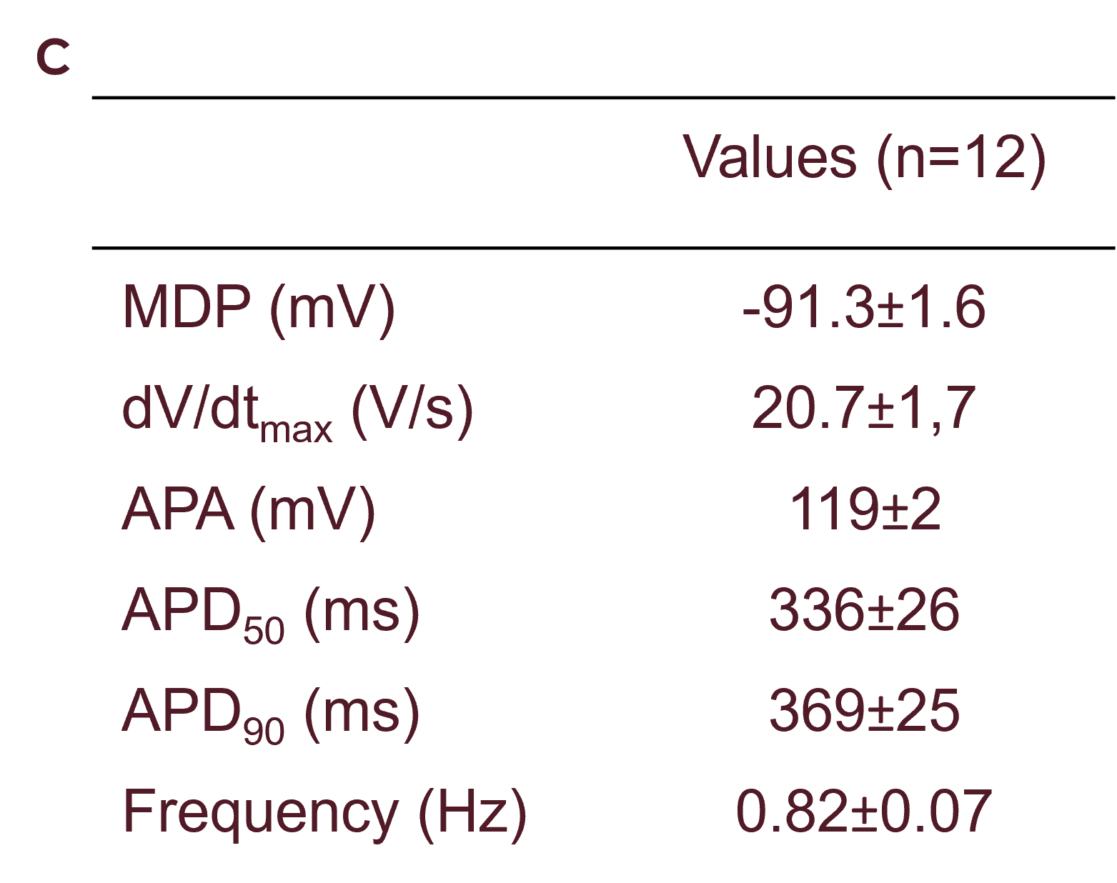 Table with averaged values of ventricular action potentials in current clamp mode. Data presented as mean ±SD (n = 12).
Ncyte® vCardiomyocytes present a relatively slow and uniform beating rate, which enables pacing for electrophysiological research.
Our scientific team is experienced in pharmacology and toxicity assays of cardiomyocytes to help you select the best drug candidates.
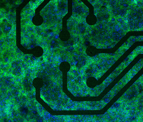 Immunofluorescence staining of Ncyte® vCardiomyocytes plated on a 96-well MEA plate: cTnT in green and DAPI in blue.
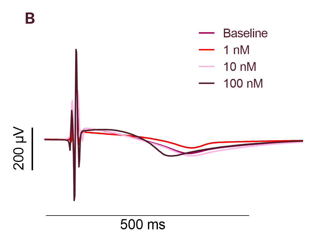 Representative MEA traces affter a 30 minute exposure to increased dosage of Isoproterenol.
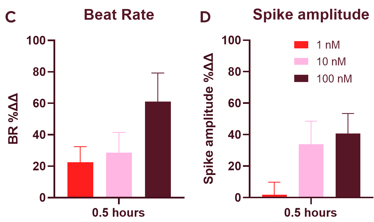 C) A 30-minute exposure to isoproterenol increased the beat rate and D) spike amplitude in Ncyte® vCardiomyocytes. Data presented as mean ±SD (n = 6) of the % change to baseline recordings for each well and time-matched 0.1% DMSO control.
Response to adrenergic stimulation with isoproterenol and ion channel blockers, nifedipine (L-type calcium channel blocker) and dofetilide (hERG channel blocker), is guaranteed for every batch.
For more data download the fact sheet
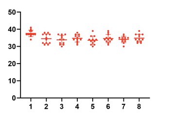 Microelectrode array baseline beating rate of a 96-well plate with Ncyte® vCardiomyocytes.
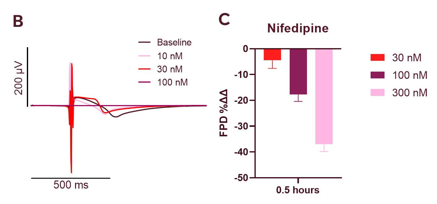 B) Representative MEA traces after a 30 minute exposure to increased dosage of Nifedipine. C) A 30-minute exposure to Nifedipine, an L-type calcium channel blocker, shortened the field potential duration of Ncyte® vCardiomyocytes in a dose dependent manner.
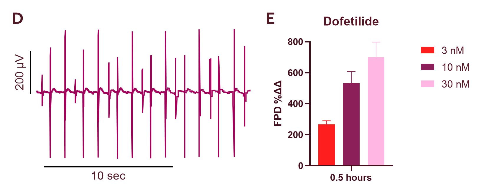 D) Representative trace after 30-minute exposure to dofetilide, a hERG (IKr) channel blocker at concentrations ≥10 nM, causes notable arrhythmias E) A 30-min exposure to Dofetilide induced a dose dependent prolongation of field potential duration in Ncyte® vCardiomyocytes. Data presented as mean ±SD (n = 6) of the % change to baseline recordings for each well and time-matched 0.1% DMSO control.
.png?width=2000&height=2000&name=nc_material%20(19).png "nc_material (19)")
Human iPSC-derived atrial cardiomyocytes
Neural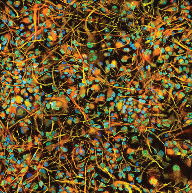
Human iPSC-derived astrocytes
Vascular
Human iPSC-derived vascular endothelial cells
Cardiac.png)
Human iPSC-derived 3D cardiac microtissue model
Neural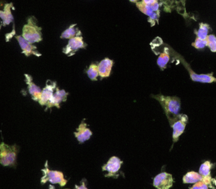
Human iPSC-derived microglia
Cardiac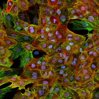
Non-Human Primate Cynomolgus iPSC-Derived Ventricular-Like Cardiomyocytes
Vascular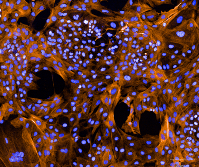
Human iPSC-derived vascular smooth muscle cells
Cardiac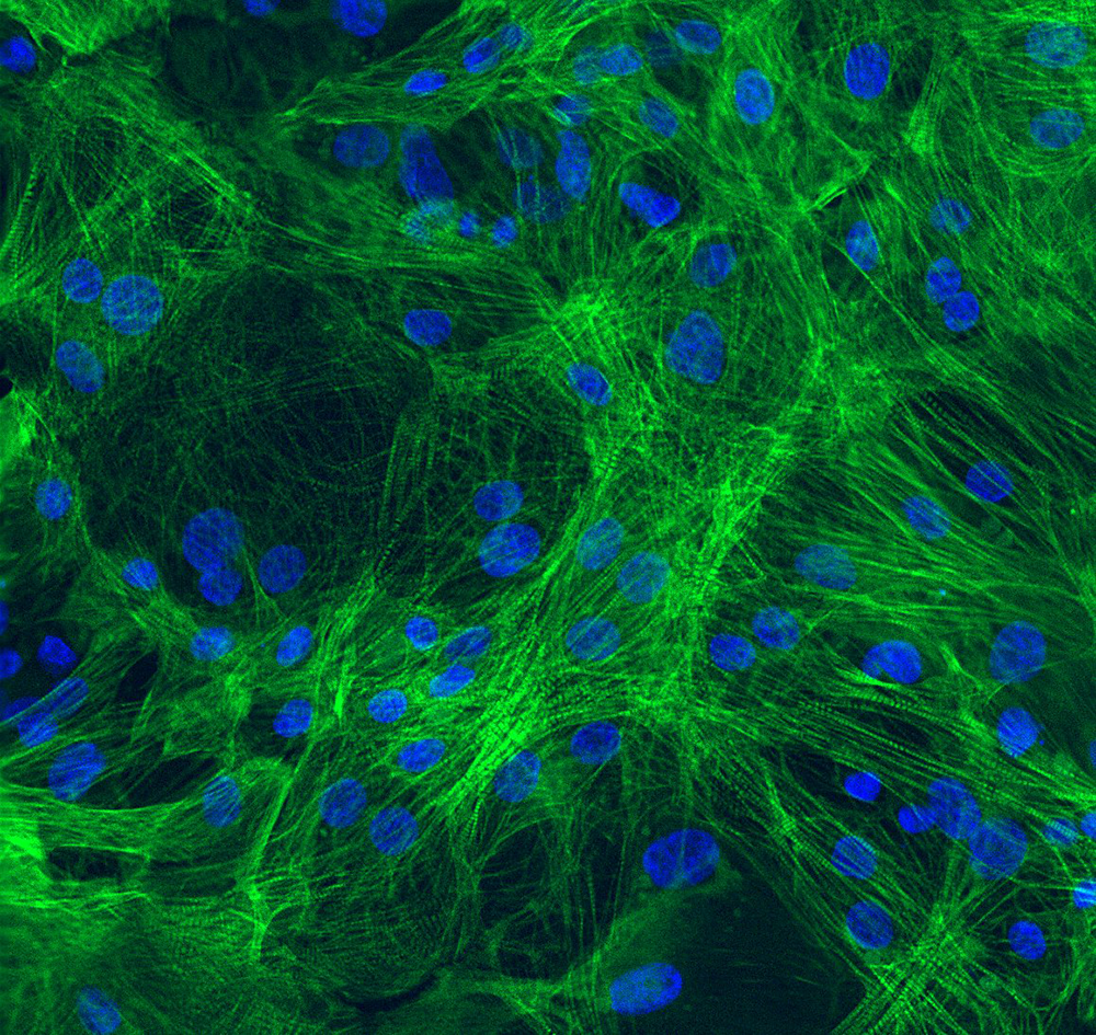
Human iPSC-derived ventricular cardiomyocytes