- High reproducibility
- Functional and physiologically relevant
- Sensitive compound-induced response
Using a standardized process, Ncardia manufactures Ncyte® Smooth Muscle Cells which are characterized with key identity markers: Calponin and α-smooth-muscle-
actin (α-SMA). For further identity verification, our scientists check the absent of CD31 expression.
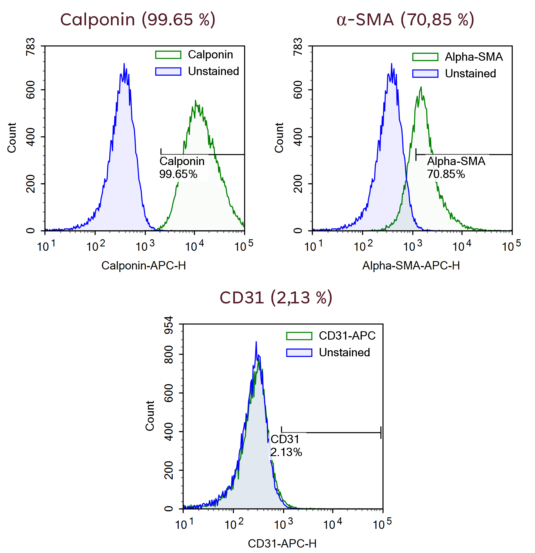 Representative histograms of flow cytometric analysis of a representative batch of Ncyte® Smooth Muscle Cells showing expression of identity markers, Calponin and α-smooth-muscle-actin (α –SMA). Bottom panel shows that CD31 (endothelial cell marker) is not expressed.
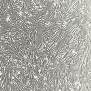 Representative bright field image of Ncyte® Smooth Muscle Cells showing cell morphology at day 7 of culture according to user guide.
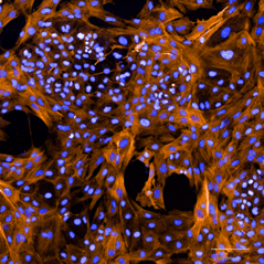
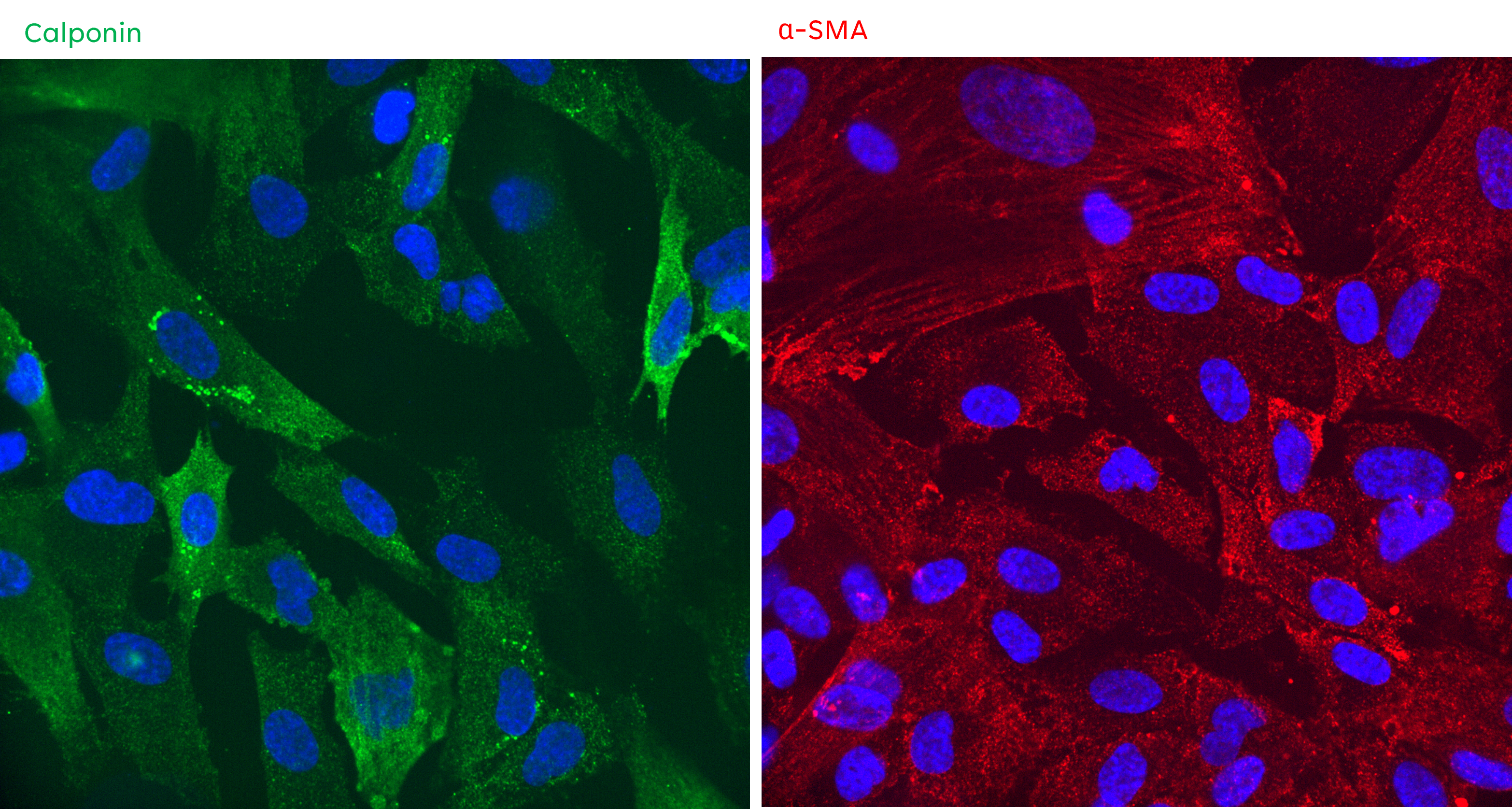 Representative images showing immunofluorescence staining of Ncyte® Smooth Muscle Cells expressing Calponin (green) and alpha-smooth-muscle-actin (red) at day 7 of culture according to user guide.
Ncyte® Smooth Muscle Cells are the ideal tool to provide relevant predictions on your drug’s effect on vaso-activity. For example, they can be used to functionally assessed calcium transients in response to compound treatment.
Our scientific team can develop a wide range of assays to evaluate the functions of your interest. As an example, the figure shows calcium transient analysis in response to endothelin-1.
 FDSS/μCELL Functional Drug Screening assessing endothelin-1 (ET-1) induced Ca2+ flux on Ncyte® Smooth Muscle Cells (cultured according to project needs). The graph shows concentration-dependent response to ET-1.
Ncyte® smooth muscle cells are a sensitive model. When used in vascular safety assays, they can produce accurate dose-dependent responses and help identify and characterize vasoactive compounds.
Our scientific team can develop assays and screenings with high reproducibility to help you make more confident decisions in early drug discovery.
 Calcium transients measured in a batch of Ncyte® Smooth Muscle Cells (cultured according to project requirements). These graphs show the expected negative response to non-vasoactive compounds like Amoxicillin and concentration-response to a vasoconstrictor compound (Angiotensin II). AstraZeneca presentation at Eurotox 2018.


Human iPSC-derived atrial cardiomyocytes
Neural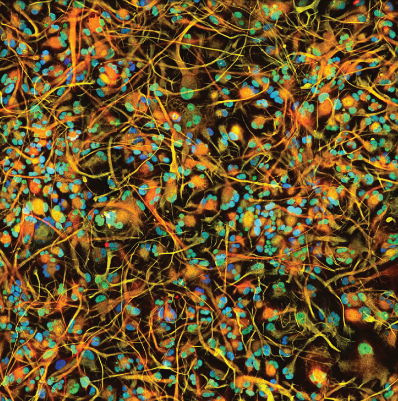
Human iPSC-derived astrocytes
Vascular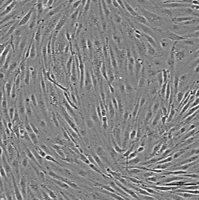
Human iPSC-derived vascular endothelial cells
Cardiac.png)
Human iPSC-derived 3D cardiac microtissue model
Neural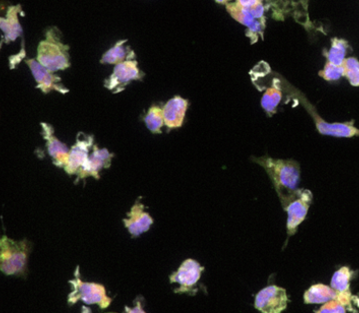
Human iPSC-derived microglia
Cardiac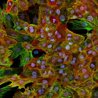
Non-Human Primate Cynomolgus iPSC-Derived Ventricular-Like Cardiomyocytes
Vascular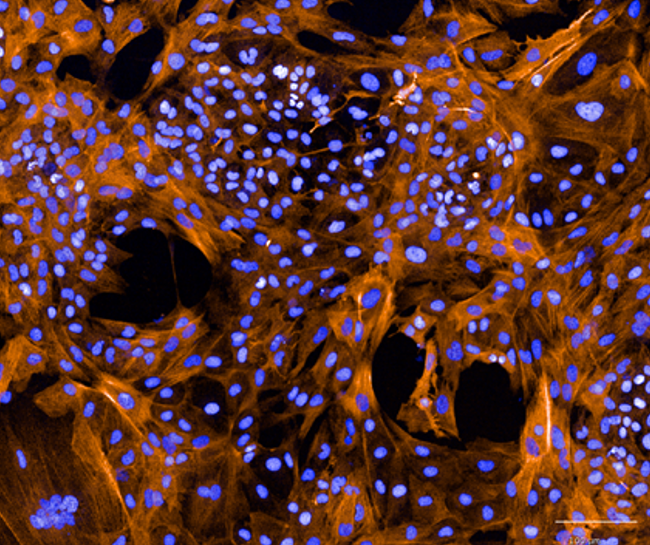
Human iPSC-derived vascular smooth muscle cells
Cardiac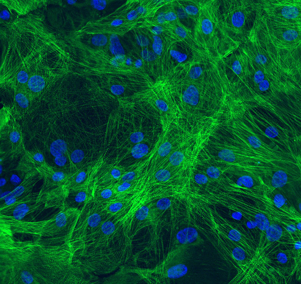
Human iPSC-derived ventricular cardiomyocytes