High-throughput screening of anti-hypertrophy drugs based on NT-proBNP biomarker detection with high content imaging
To identify new treatments for cardiac hypertrophy, our scientists generated an iPSC-derived model of hypertrophic cardiomyopathy and used HCI for hit validation with 341 compounds. The intracellular expression of NT-proBNP biomarker was used as readout to determine the efficacy of the anti-hypertrophic hit compounds.
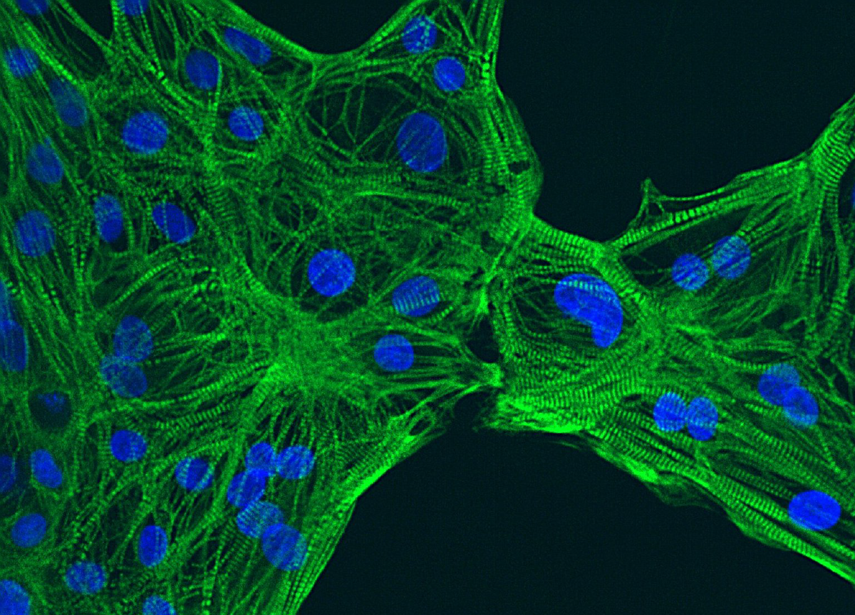
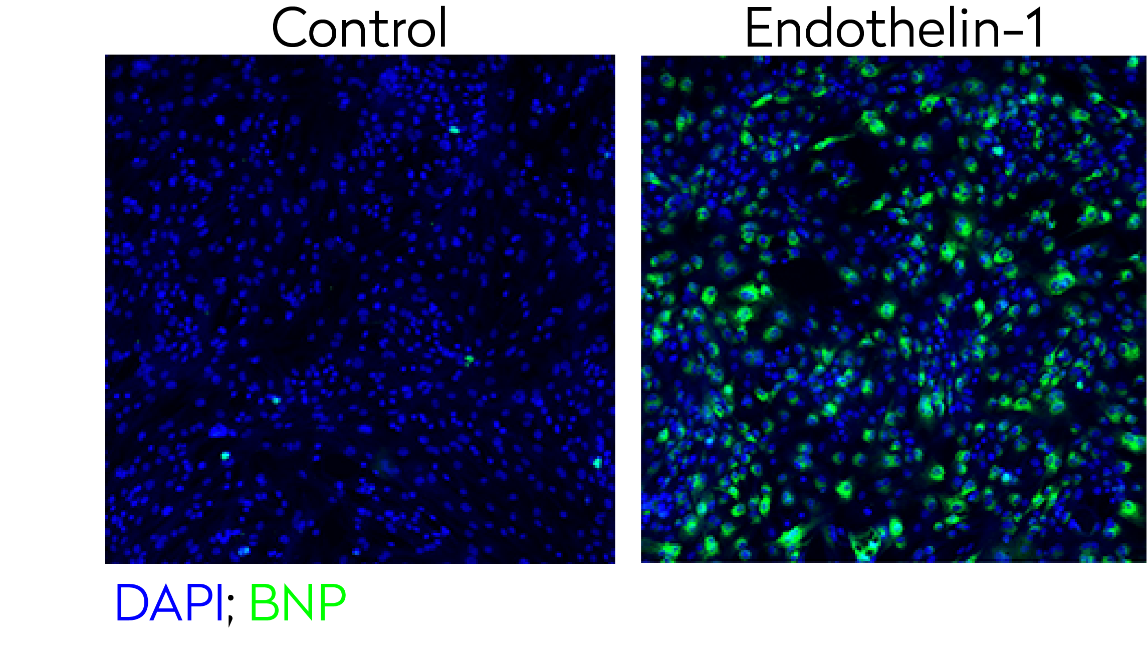 Representative confocal images of control and endothelin-1 treated human iPSC-derived cardiomyocytes stained for BNP hypertrophy biomarker (green) and DAPI (blue - nuclei).
Evaluation of lysosomal dysfunction in a human iPSC-derived model of Alzheimer’s disease with high content imaging.
Lysosomal dysfunction is a hallmark for neurodegeneration. Using HCI, we can detect lysotracker marker to localized lysosomes and determine the capacity of new therapeutic candidates to rescue lysosomal dysfunction.
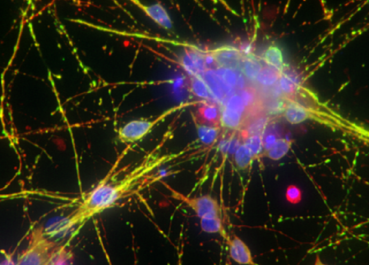
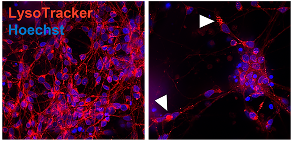 Representative confocal image of control and AD iPSC-derived neurons showing lysosomal distribution and pattern (Lysotracker, red). Arrowheads point to lysosome accumulation in the cytoplasm.
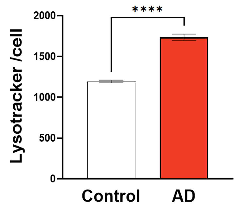 Graph showing quantification of Lysotracker cell average intensity (RFU) in control and AD iPSC-derived model
Study compound-induced effects on blood vessel formation
Assay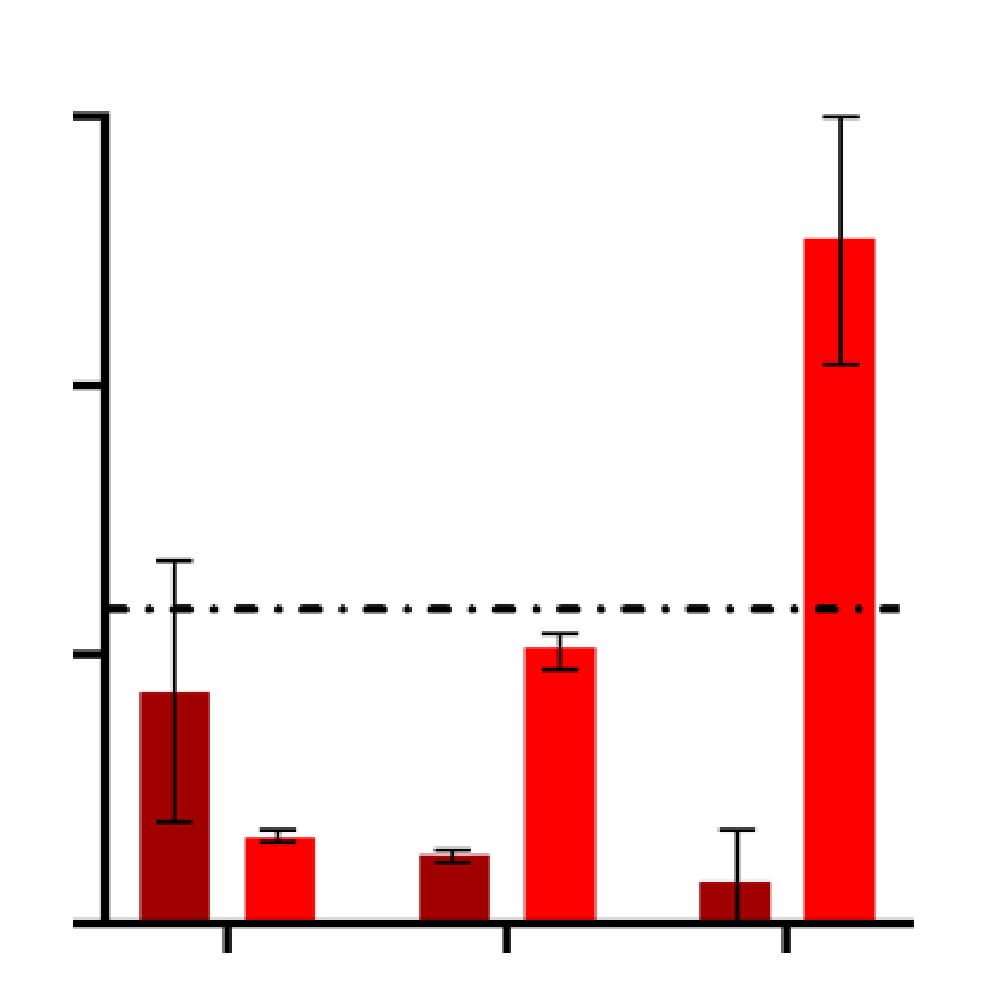
Evaluate and quantify the levels of clinically relevant biomarkers
Assay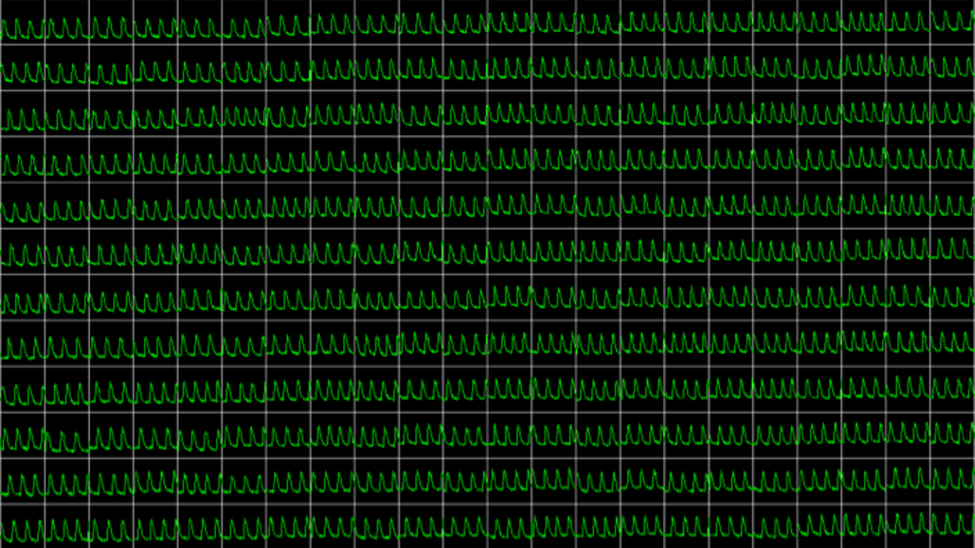
Obtain real-time recordings of intracellular calcium fluctuations
Assay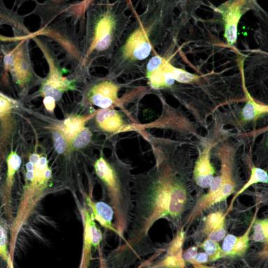
Get in-depth and unbiased insights into the effects of therapeutic candidates on cells
Assay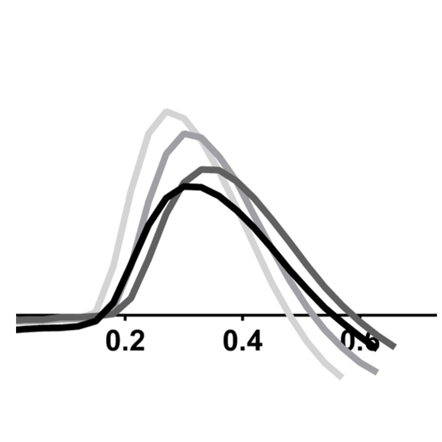
Study drug-induced effects on contractility of cardiac or skeletal muscle cells
Assay
Determine the electrophysiological effects of your therapeutic candidates
Assay
Evaluate endothelial permeability in short and long term
AssayStudy drug-induced phenotypic changes in the cell model of your interest
Assay
Assess functional toxicity across multiple human iPSC-derived tissues in a single integrated workflow to support safer IO development decisions.
Assay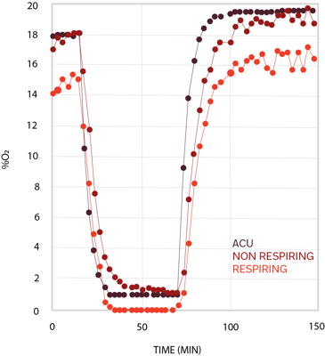
Obtain precise information on compounds' impact on metabolic processes